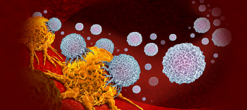
Brief Overview of Clonality Analysis and Its Applications

Clonality analysis is a technique used to determine whether a population of cells is derived from a single cell or multiple cells. It is used in various fields such as cancer research, immunology, and microbiology. Clonality analysis can be performed using various methods such as PCR-based methods, flow cytometry, and next-generation sequencin.
In cancer research, clonality analysis can be used to identify the origin of tumors and to detect clonal diversity in tumors1. In immunology, clonality analysis can be used to study the development of B and T cells and to identify monoclonal antibodies. In microbiology, clonality analysis can be used to study the evolution of bacterial populations.
Clonality analysis can be performed using various methods such as PCR-based methods, flow cytometry, and next-generation sequencing. Clonality assessment makes use of the feature that clonal populations of cells have identical molecular footprints of the antigen receptor encoded by the IG genes. Clonality analysis can be used to identify the origin of tumors and to detect clonal diversity in tumors2. In immunology, clonality analysis can be used to study the development of B and T cells and to identify monoclonal antibodies. In microbiology, clonality analysis can be used to study the evolution of bacterial populations.
Clonality analysis has several advantages. It can be applied to all tumor sources, including archival tissues (FFPE) and has good sensitivity and excellent tumor specificity allowing. Clonality testing uses this feature and facilitates the discrimination between clonally expanded cells and reactive cells with diverse IG and/or TR rearrangements. NGS-based clonality analysis has high sensitivity and possibilities for accurate clonal comparison.
Clonality analysis service using FISH (Fluorescence In Situ Hybridization) is a technique used for biological research to detect chromosomal abnormalities and gene rearrangements in cells. It is used to identify the origin of tumors and to detect clonal diversity in tumors. Creative Bioarray offers Clonality Analysis Service (FISH) for scientific research as follows: Probe design, Probe synthesis, Chromosome spread preparation, FISH on chromosome spreads, Imaging, Data analysis (karyotyping and integration pattern analysis).
Recent developments such as single cell sorting and high-throughput imaging allow for the acquisition of evidence supporting clonality. Rederiving a clonal cell line by additional limited dilutions is time-consuming, costly and, crucially, may affect production and growth rates of cell lines. This is especially undesired when time lines for submission are short. Creative Bioarray provides the genetic characterization of (CHO) producer cell lines by FISH, which offers information on transgene integrity and integration sites.
The steps for developing a FISH probe:
Target selection: This is a critical step. Keep in mind that FISH can effectively locate chromosomal regions of more than several thousand basepairs.
Probe design: The probe should be designed to be specific to the target sequence.
Probe synthesis: The probe is synthesized with a fluorescent molecule attached to it.
Probe labeling: The probe is labeled with a fluorescent molecule.
Probe purification: The probe is purified to remove any unincorporated fluorescent molecules.
Hybridization: The probe is hybridized to the target sequence.
Washing: The unbound probe is washed away.
- Like
- Reply
-
Share


About Us · User Accounts and Benefits · Privacy Policy · Management Center · FAQs
© 2024 MolecularCloud